





Question 58 · LDEK · Access and canal preparation
Which clinical situation does the following X-ray show?
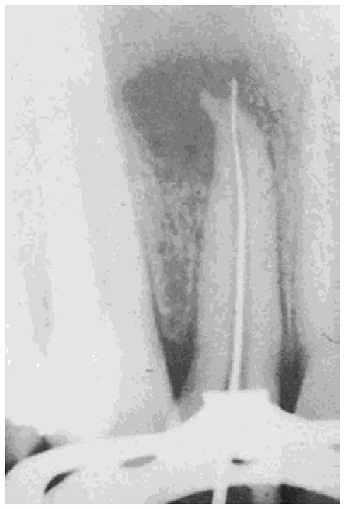
- A. performing recapitulation.
- B. extrusion of gutta-percha cone beyond the apical constriction.
- C. checking patency of the root canal after correct orifice preparation.
- D. insertion of a too narrow endodontic file into the root canal during its preparation.
- E. creation of an apical perforation during endodontic treatment.
Hint
Compare the width of the canal lumen with the width of the instrument and check whether the file touches the canal walls.
The answer, the explanation and a note on every option are available once you log in. The account is free: topic tests, a mock exam, mistake review and statistics.
More questions on Conservative dentistry
- A 35-year-old patient complains of pain to mastication on teeth 15 and 16. Percussion test is positive (patient reacts with pain). Clinical and…
- Indicate which irrigant can replace EDTA and sodium hypochlorite to remove the smear layer:
- Which of the following is the first, clinically visible sign of the carious lesion:
- Indicate the false statement concerning dental erosion:
- Indicate the true statements concerning the process of composite polymerization:
- In which direction the cut is made in the procedure of hemisection of mandibular molars?